SELLS Animal Health Update April 2021
April 2021
Download Pdf (1,032kb)
Monthly disease survey results
Alex Stephens, Yass.
Your local District Vet can help you to investigate, diagnose and manage herd health or stock death issues in your herd or flock. We provide impartial advice and can assist you with disease management and your biosecurity plan. Each month we provide this report of diseases and issues detected and managed in the last month by producers, their veterinarians and animal health advisors.
Sudden death in pigs: African Swine Fever (ASF) and Classical Swine Fever (CSF) were tested for and ruled out in a suspicious case of sudden death of two weaner grower pigs. Thanks to the private vet who identified the potential risk and alerted us. A conclusive diagnosis was not reached, but the deaths were most likely due to salt toxicity from accidental water deprivation.
Flystrike: Recent heavy rains followed by a week of warm weather has caused continued flystrike issues in sheep, and work for producers. To prevent losses, you need to keep an eye on your stock, treat affected stock and have a plan to prevent and manage future episodes. If left unchecked Flystrike can become a serious animal welfare issue, producers have been reported to the RSPCA about this in the past.
Heavy worm burdens causing deaths: Haemonchus contortus (barber’s pole worm) has been killing mature sheep and hoggets in all areas of the South East. Conditions have been perfect for barber’s pole and scour worm numbers to increase rapidly. Deaths have been seen when sheep have been drenched with an ineffective drench, particularly abamectin, or not rotated to clean pastures after drenching. Drench resistance is common, know which drenches are effective on your property and test your sheep now with a faecal egg count (FEC) to avoid losses.
Persistent scouring and weight loss in hoggets: A case of persisted scouring was investigated and found to be a bacterial scour caused by a combination of yersinia and salmonella bacteria. The period of cold wet weather and stress could have contributed to this outbreak. The diarrhoea persisted despite drenching, the right treatment was only possible after the issue had been diagnosed by a vet.
Footrot inspections: Inspectionshave continued from traces, saleyard detections and lameness investigations, with several new properties being issued a biosecurity direction to contain and resolve virulent footrot on their property. Virulent footrot is a notifiable disease. This means that if you suspect virulent footrot in your flock you must notify your District Veterinarian. The mild wet autumn following a wet summer has ensured we have a challenging footrot season in front of us for benign and virulent footrot. Foot bathing and movement of sheep to drier paddocks are important in managing the disease. You can read more about footrot in Evelyn Walker’s article below.
Foot abscess in rams: As joining commences or finishes, there have been many calls and visits to rams with sore feet. Footrot must be ruled out but in most cases the lameness has been caused by foot abscess. Foot abscess is very painful, with swelling, redness and pus. Inspect rams at least 3 months before joining and again at each month up to joining to ensure they have four sound feet well before joining. Continue to inspect them during joining and consult with your private vet to ensure rams get the treatment they need.
Pinkeye: Outbreaks have been seen in several mobs of sheep. Pinkeye in sheep can be caused by several organisms, which are most likely being transferred between sheep by small flies. Although the lesions will heal in most cases without treatment, sheep can become temporarily blind in both eyes, get injured and die. Ewes in the last few weeks of pregnancy may be at risk of metabolic diseases if blindness stops them from grazing high quality feed. Although yarding sheep can cause further transmission of pinkeye it is best to recognise and treat affected mobs sooner rather than later, by first checking for grass seeds then using a treatment such as terramycin spray. Orbenin eye ointment works sometimes, but not always, due to the variety of organisms that can cause the issue. Talk to your private veterinarian about treatment options. In some cases, injectable antibiotics may be required.
Photosensitisation caused by hairy panic: Hairy panic associated photosensitisation (sensitivity of skin to light)has been seen in flocks on the Monaro. Hairy panic is a summer perennial grass which is more common in the South East due to summer rainfall. The first symptoms noticed are swollen ears, eyelids and noses. Affected animals should be removed from the grass to avoid severe skin damage or long-term liver damage. Talk to your vet or agronomist about safer grazing strategies.
Nasal bots: In sheep, nasal bots have been discovered during a post-mortem exam where the cause of death was anaemia caused by barber’s pole worm. Nasal bots are the large maggot of the sheep nasal bot fly. They usually cause little harm but live inside the nasal passages but in some cases such as this one, they cause nasal irritation and consequently a persistent nasal discharge. Most drenches containing an ML such as ivermectin will kill nasal bots.
ILT was diagnosed in a small backyard poultry flock: Infectious laryngotracheitis (ILT) is a notifiable and economically important respiratory disease of poultry. It is highly contagious and easily transmitted through infected birds, faeces or anything that has been in contact with birds. It causes severe difficulty breathing, noisy respiration, coughing and death but can also cause chronic conjunctivitis and nasal discharge. Be careful when introducing birds to your flock or showing birds book a consult with your private vet or contact your District vet if you are seeing respiratory symptoms or deaths.
District Vet conference: The 102nd District vet conference was held at Broken Hill from 23 to 25 March and four of the South East district vet team made the journey to attend. The annual DV conference dates back to the “stock inspector” days. Held in a different Local Land Services office, or DPI laboratory location each year, it gives the opportunity to share experiences of disease investigations, surveillance, emergency response and animal health advisory work with our colleagues. We hear about the differences and similarities of the many farming landscapes and farmers across our state. It also fosters a strong collaborative relationship with Department of Primary Industries (DPI) and State Veterinary Diagnostic Lab (EMAI) staff. Local Land Services and DPI were tremendous supporters of this conference and we were joined by Local Land Services Board Chair Richard Bull, Local Land Services CEO David Witherdin, NSW Chief Veterinary Officer Sarah Britton and Director of the Elizabeth Macarthur Agricultural Institute (EMAI) Jim Rothwell.
Fluke treatment options
Lou Baskind, Queanbeyan-Palerang
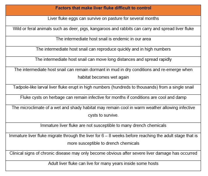
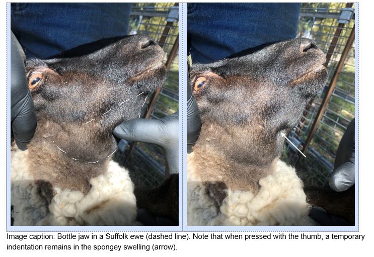
Liver fluke is found on the tablelands and much of the coastal areas of eastern NSW. In wet years, the distribution of liver fluke can spread westwards. The parasite infects a range of mammal hosts and causes liver damage and blood loss. The result of the infection ranges from initially undetected production losses (reduced growth rates, wool growth and fertility) to observable clinical signs such as ill-thrift, “bottle jaw”, pale or yellow membranes in mouth and eyes, to death. Liver fluke control requires an understanding of the complex lifecycle of the liver fluke parasite, including the intermediate host snail’s habitat requirements.
The liver fluke parasite lifecycle
Liver fluke eggs develop through a complex lifecycle, via an intermediate host snail, eventually becoming infective cysts that are then eaten by the mammal host. In Australian agriculture our most important host species are cattle, sheep, goats and alpacas but a range of other animals can carry and spread the fluke parasite including deer, pigs, goats, kangaroos and wallabies, and rabbits. The broad host range which includes wild and feral animals rules out the complete eradication of liver fluke.
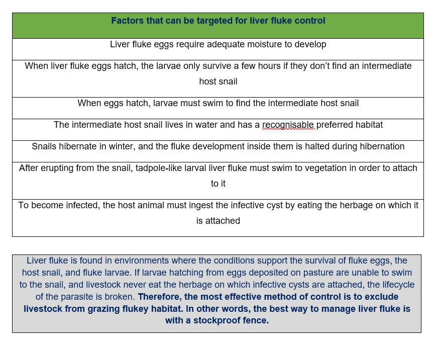
Liver fluke eggs are shed in the dung of the host animal onto pasture. These eggs are extremely robust and can survive on pasture for several months. Once deposited on pasture the eggs await appropriate conditions – adequate moisture and temperatures above 10◦C. They develop for several weeks with their development more rapid in warmer weather. Once hatched, larvae swim through water to find an intermediate host, a freshwater snail called a lymnaeid snail. The larvae can survive only a few hours between hatching from the egg and burrowing into a snail. Once within the snail the larvae multiply. This takes from one to three months, again developing faster in warmer weather. Now grown into a tadpole-like shape, the larvae erupt in their hundreds from the snail. They swim to vegetation and attach, then form a tough protective casing. This is the infective stage, the “infective cyst”.
A host animal eats the infective cysts as it grazes normally. Once inside the intestine of the host, the immature liver fluke hatch from their tough casing and burrow through the intestinal wall towards the liver. For six to eight weeks immature liver fluke migrate through the liver while feeding on the liver tissues and blood cells. This causes substantial damage. Production losses from this damage will be significant, even if clinical signs are not observed by the livestock manager.
Eventually the liver fluke enter the bile ducts - small tubes in the liver that carry liquid bile from the liver to the gallbladder and then to the intestine. Once in the ducts they double in size, become mature and start laying eggs that travel from the liver in the bile and are shed in the dung of the host.
The intermediate host snail
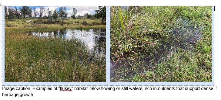
The intermediate host snail is a freshwater snail that lives in slow flowing or still waters at the edge of springs, small creeks, dam inflows and outflows, irrigation channels, poorly drained drainage channels and water trough surrounds. The snails prefer waters that are rich in nutrients and support dense herbage growth. These areas are often referred to as “flukey”.
The snails reproduce by laying about 3000 eggs per month. It takes approximately one month for an egg to develop through to an adult snail able to lay the next generation of eggs. In winter, the reproduction rate is much slower and in very cold conditions the snails hibernate. The snail can move long distances, even against the water current, meaning it can spread rapidly.
In periods of hot and dry conditions, the snails can remain dormant in dry mud and quickly return to normal activity when wet conditions return.
Talk to the Natural Resource Management team at your nearest Local Land Services office about funding opportunities for keeping stock out of waterways.
Other strategies
In practice, fencing out flukey habitat takes time and money and will be part of a longer-term whole farm plan. Also, the feed available in flukey habitat may be needed, especially where it provides high quality feed when there is not much else on the farm. So other management strategies must come into play.
Grazing with less susceptible stock
Minimise grazing of flukey habitat by the most vulnerable livestock: young cattle, sheep, goats and alpacas. After being infected by liver fluke, adult cattle develop a resistance to subsequent infection due to liver scarring (fibrosis) that forms a physical barrier to fluke migration. The cattle survive and do not show overt disease. Their livers are large, and they retain enough undamaged liver to function. Note that liver function may be permanently reduced in some cattle with long term reduced productivity.
Sheep, goats and alpacas don’t develop resistance to liver fluke so can suffer at any age. Alpacas are thought to be particularly susceptible to liver fluke, perhaps as their liver is small and therefore damage is more rapidly overwhelming. Alpacas are also prone to spread of bacteria, such as e. coli, from the damaged liver to the heart, which can cause endocarditis and heart failure.
Chemical drenches to reduce infection levels
Strategic chemical treatments are used to reduce the liver fluke population, but they cannot be used alone to eliminate liver fluke in areas where it is common. Chemical drenches are used once to three times per year depending on how badly properties are affected and how susceptible the livestock are. In south eastern Australia, the treatment given in early winter (April/May) is the most important as this represents the time of highest liver fluke levels. An early spring treatment and a summer treatment can also be given. Speak to an advisor or your local District Veterinarian to discuss requirements for chemical treatments on your farm.
Grazing rotation with chemical drenches
If flukey areas must be grazed, consider giving an effective fluke drench two weeks before moving stock into that area. This prevents stock from shedding fluke eggs while grazing there. Try to restrict this grazing period to less than six weeks, moving stock back to non-flukey country before they are shedding eggs in their dung again.
Quarantine drenching
When buying in livestock, treat them with a combination drench effective against all lifestages of liver fluke, then hold them in non-flukey paddocks for 21 days before testing for liver fluke to ensure the treatment was effective. Speak to your advisor or local District Veterinarian about testing options.
Chemical drenches
At the early winter drench in April/May, animals that have grazed flukey areas through spring and summer will be host to early immature, immature and adult-aged liver flukes. A drench that eliminates all these life stages should be used.
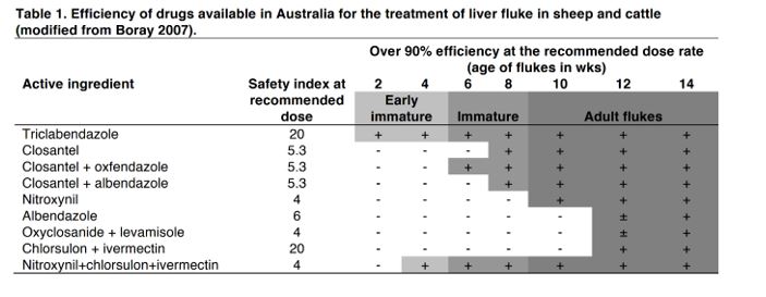
Triclabendazole is the chemical of choice for the early winter drench for sheep and goats. Either triclabendazole, or a nitroxynil and clorsulon combination is the drench of choice for the early winter drench for cattle.
Overuse of chemical drenches should be avoided as resistance is likely to develop. To slow the development of resistance a different chemical should be used for the spring and summer treatments if they are required.
Drenching alpacas
While alpacas are particularly susceptible to liver fluke, and require chemical drenching just as other livestock do, no chemical products are currently registered for alpacas for treatment of liver fluke. Consult with your private veterinarian about options for an off-label authority. An off-label authority allows a registered veterinary chemical to be used in a manner that is not specified on the product label. Alpacas require different doses to those of sheep and cattle for the chemical to be effective. Private veterinarians are legally required to examine the alpacas once or twice per year to supply the off-label authority.
Recognising the disease
Liver fluke disease is the result of tissue damage, thickening of the bile ducts and blood loss. If very large numbers of immature liver fluke migrate at once, the tissue damage and blood loss will quickly overwhelm the host animal, causing rapid death.
This acute form of disease may be accompanied by signs of pain such as tucking up in the abdomen and reluctance to move, anaemia which is seen as pale membranes of the mouth and eyes, or jaundice which may cause the membranes or the skin to appear yellow.
Sudden death may also be the result of Black Disease in animals that are not up to date with clostridial vaccinations. The damage to the liver caused by migrating immature liver fluke can activate a bacterium called Clostridium novyi, which produces a fatal toxin.
If the immature liver fluke numbers are somewhat less, the clinical signs include anaemia, jaundice and failure to thrive, with animals potentially dying after several weeks.
In the chronic form of disease, clinical signs come on slowly, and a “bottle jaw” develops. This form of the disease is most typical in autumn and winter when animals have been eating infective cysts over the course of spring and summer. In sheep it can be difficult to distinguish chronic liver fluke disease from barber’s pole worm infection.
Look out, Footrot about
Evelyn Walker, South Coast
Footrot detections in sheep are on the rise in the Shoalhaven district. Footrot is a serious highly contagious disease of sheep and goats caused by the bacteria, Dichelobacter nodosus.
This year’s mild temperatures and rainfall over spring, summer and into autumn created the ideal environment for the footrot bacteria to show itself in both sheep and goat feet. Footrot does not flourish under dry, hot environmental conditions, so we see fewer cases in times of drought. Footrot thrives in warm moist conditions. For an in-depth look at footrot in sheep and goats, check out the NSW DPI Footrot Primefact.
Whether you have small or large numbers of sheep or goats—no individual or breed is immune to footrot. Virulent footrot in sheep and goats is notifiable in NSW as per schedule 1 of the Biosecurity Regulation 2017, This means you must report any suspicion to your nearest Local Land Services District Veterinarian. See contact details for your local District Veterinarian below. Footrot creates a biosecurity impact, and it is your biosecurity duty to eliminate or minimise the risk and impact of footrot on your property. Failure to report is an offence under Section 23 of the Biosecurity Act 2015 and could result in hefty penalties.
It's best practice to check your stock daily and have any signs of lameness investigated by your veterinarian. There are many causes of lameness and symptoms can range from a mild limp in one or more legs to non-weight bearing or preferring to lie down more than usual. You might observe pressure sores on their knees or chest from prolonged periods of rest. When observing your sheep or goats, catch sheep and have a close look at their feet. With such a prolonged wet year sheep or goat’s feet did not wear down naturally, resulting in a lot of overgrown hooves that need to be paired. Overgrown heels and toes of the hoof affects mobility and foot health. It can be painful depending on the inciting cause, and pockets that form can serve as a niche hide away for bacteria. Regular pairing of the feet will improve overall foot health and optimise air flow. Cull those with consistently bad feet.
In addition to maintaining foot health with regular trimming, pay close attention to the health of ram and buck feet before joining, ensuring they are 100% sound. The practice of lending and hiring out breeding stock should be avoided due to the risk of spreading footrot and/or other diseases.
Minimise your risk by having a biosecurity plan in place for all incoming stock. In addition to due diligence checks, record checks (e.g. NVD, Animal Health Declarations), prophylactic drench and vaccination treatments, you should isolate new stock for a minimum of 4 weeks and ideally until after a footrot spread period. Beware the footrot bacteria can lie dormant within the foot and only show itself when the conditions are just right. If the isolated animals break down with footrot, you haven’t compromised the rest of your flock.
If you are a small landholder, don’t let limited paddock space or size cause you to skimp out on biosecurity. With careful planning, you can work out a quarantine plan that suits your needs.
For questions on footrot, biosecurity planning, or animal husbandry in general, drop in or ask to speak to one of friendly Biosecurity Officers, Agricultural Advisors or District Veterinarians.
National Livestock Identification System
Michelle Meyers, 4th Year University of Sydney Veterinary Student
Australia relies on The National Livestock Identification System (NLIS) for the identification and traceability of individual livestock. The NLIS ensures biosecurity and food safety is maintained at high standards to provide quality assurance to the market. Australia was the first country to initiate a livestock tracing system, given the high number of international visitors and trading partners. The NLIS compiles animal identification and property identification onto a web database to assist in the traceability of all stock. Each individual animal is legally required to keep the same identification number for the entirety of its life. These strict measures are in place to ensure that a life history of movements is traceable in the event of disease incursion or in a situation of a contaminant. The Biosecurity Act 2015 governs the NLIS system in NSW.
To properly use NLIS, a property identification code (PIC), national vendor declaration (NVD), and quality record keeping are considered essential. A property identification code is an 8-character code arranged by each state or territory used to classify livestock-producing properties. The NVD is a legal document used to identify food safety concerns and any treatment history.
Electronic radio-frequency identification devices (RFID/NLIS tags) are used to maintain the traceability of cattle. These devices can range from a visual ear tag, rumen bolus, or a combination of both. The color of the devices varies, where breeder devices are white and post-breeding devices are orange. If a rumen bolus is used, the same coloring system applies using a large permanent ear tag.
Under the NLIS regulations all identifiable stock (Cattle and Sheep) must have an NLIS tag attached prior to leaving their property of birth. It is the responsibility of the owner or person in charge of the stock and the transporter to check stock prior to it leaving the property. Movement of livestock must be entered into the NLIS database within 48 hrs. For private sales, the buyer or receiver must record these movements.
Maintaining Australia’s food safety reputation is critical. Through the assistance of the NLIS, tracing stock from the property of origin to the point of sale provides clear evidence for the integrity of all products sold throughout Australia.